
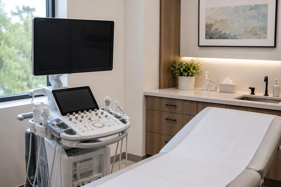






Medical imaging is an important part of modern healthcare. It enables healthcare professionals to examine structures inside the human body without always needing surgery or other invasive procedures. Through imaging technologies such as X-rays and ultrasound, clinicians can investigate symptoms, assess injuries, monitor certain medical conditions and make informed decisions about patient care.
At Bethwell Imaging Centre, we recognise the importance of evidence-based practice in supporting accurate assessment and responsible healthcare decisions. Medical imaging provides valuable information that, when combined with a patient’s medical history, physical examination and other relevant investigations, helps healthcare professionals develop appropriate care plans.
Understanding medical imaging can help patients appreciate why their doctors request particular examinations, how these procedures are performed and what they should expect before, during and after an examination.
1. What Is Medical Imaging?
Medical imaging refers to the use of specialised technologies to produce images of organs, tissues, bones and other structures inside the body. These images help healthcare professionals investigate medical concerns that may not be fully understood through a physical examination alone.
For example, a doctor may request an X-ray when a patient has a suspected bone fracture. An ultrasound examination may be requested to assess certain abdominal organs, monitor a pregnancy or investigate a lump.
Medical imaging does not replace clinical assessment. Instead, it provides additional information that can help healthcare professionals understand a patient’s condition and determine the most appropriate next steps.
The choice of imaging examination depends on several factors, including the symptoms, the suspected condition, the body part being examined, the patient’s medical history and the clinical question that needs to be answered.
2. Common Types of Medical Imaging
Different imaging technologies serve different purposes. Understanding their uses helps explain why one examination may be recommended over another.
A. Digital X-Ray
Digital X-ray uses a small amount of ionising radiation to produce images of structures inside the body. It is commonly used to examine bones and certain parts of the chest.
Healthcare professionals may request an X-ray to:
- Investigate suspected bone fractures and dislocations.
- Assess certain chest conditions, including suspected pneumonia.
- Examine some joint and bone abnormalities.
- Monitor selected conditions or assess progress after treatment.
Digital X-ray systems capture images electronically, allowing them to be viewed on a computer and stored digitally.
Although X-rays are useful diagnostic tools, they involve radiation exposure. Healthcare professionals should ensure that each examination is justified and performed using appropriate radiation-safety measures. Patients should inform the healthcare team if they are pregnant or think they may be pregnant.
B. Ultrasound Scans
Ultrasound uses high-frequency sound waves to produce images of structures inside the body. Unlike X-rays, it does not use ionising radiation.
Ultrasound examinations may be used to:
- Examine certain abdominal organs, including the liver, kidneys and gallbladder.
- Assess the uterus and ovaries.
- Monitor pregnancy when clinically indicated.
- Investigate selected lumps, swellings and soft-tissue concerns.
- Guide certain medical procedures.
The type of ultrasound examination determines how a patient should prepare. For some abdominal scans, fasting may be necessary, while some pelvic examinations require a full bladder. Other examinations may not require special preparation.
Patients should follow the instructions provided when their examination is scheduled rather than assuming that every ultrasound requires the same preparation.
C. Doppler Ultrasound
Doppler ultrasound is a specialised technique that assesses blood flow through blood vessels. It can help healthcare professionals evaluate the direction and characteristics of blood movement.
Depending on the clinical concern, Doppler studies may be used to investigate selected vascular conditions, assess blood flow in particular vessels or evaluate circulation in certain clinical situations.
The healthcare professional requesting the examination determines whether a standard ultrasound, Doppler study or another investigation is appropriate.
D. Other Imaging Technologies
Other medical imaging technologies include computed tomography (CT) and magnetic resonance imaging (MRI).
CT uses X-rays to create detailed cross-sectional images of the body. MRI uses strong magnetic fields and radio waves to produce detailed images of organs and tissues.
These technologies may be useful when more detailed information is required. Their availability and suitability vary between healthcare facilities and depend on the patient’s condition.
Patients should consult their healthcare professionals about the most appropriate investigation for their particular needs.
3. Why Is Medical Imaging Important in Healthcare?
Medical imaging contributes to healthcare in several important ways.
A. Supporting Diagnosis
Many medical conditions produce symptoms that overlap with those of other illnesses. Pain, swelling, fever and difficulty breathing, for example, may have different causes.
Imaging can provide additional evidence to help healthcare professionals investigate these symptoms. A chest X-ray may help assess a suspected lung condition, while an ultrasound may help investigate certain abdominal complaints.
Imaging findings must always be interpreted alongside the patient’s symptoms, medical history, examination and other relevant test results.
B. Detecting Injuries and Abnormalities
Some injuries and abnormalities cannot be adequately assessed through physical examination alone. Imaging may help identify fractures, certain organ abnormalities, fluid collections or other findings that require further investigation.
The information obtained can guide decisions about additional tests, referrals or treatment.
However, not every condition is visible on every type of scan. A normal imaging result does not necessarily rule out all possible causes of a patient’s symptoms.
C. Guiding Treatment Decisions
Medical imaging helps healthcare professionals choose suitable management options by providing information about the location, size or characteristics of certain abnormalities.
For example, imaging may help a clinician determine whether a suspected injury requires further assessment or whether a particular finding needs specialist review.
Treatment decisions should not be based on images alone. They should consider the patient’s overall health, clinical findings, preferences and the benefits and risks of available options.
D. Monitoring Selected Conditions
Some medical conditions require follow-up examinations to assess changes over time. Depending on the condition, imaging may help healthcare professionals determine whether an abnormality has changed or whether further investigation is needed.
Repeated imaging should be requested when clinically appropriate, taking into account the benefits of obtaining new information and any associated risks, including radiation exposure where applicable.
E. Supporting Preventive Healthcare
Medical imaging can contribute to preventive healthcare when used as part of an appropriate screening or assessment programme.
For example, selected breast imaging examinations may form part of breast cancer screening or the investigation of breast symptoms. Other imaging examinations may be recommended for specific individuals based on their health needs and risk factors.
It is important to understand that not every imaging examination is a screening test. Patients should seek professional advice about which screening services are appropriate for their age, medical history and individual risk.
4. How Medical Imaging Improves Patient Care
The value of medical imaging extends beyond producing pictures of the body. It can improve the information available to healthcare professionals and help patients understand their care.
Better-informed clinical decisions: Imaging findings can help clinicians evaluate possible causes of symptoms and decide whether additional investigations or referrals are needed.
More targeted investigations: When a clinician has a clear clinical question, an appropriate imaging examination may provide useful information without resorting to unnecessary or unsuitable tests.
Improved communication: Imaging reports and, where appropriate, the images themselves can help healthcare professionals communicate findings and explain the next steps in a patient’s care.
Support for ongoing care: Follow-up imaging may help assess selected conditions over time and contribute to decisions about further management.
Patient reassurance through explanation: Clear communication about the purpose, process and findings of an examination can reduce uncertainty. Nevertheless, healthcare professionals should avoid promising that an examination will always provide a definitive answer.
These benefits depend on selecting the right examination, obtaining images of suitable quality, interpreting the findings appropriately and communicating the results responsibly.
5. What Should Patients Expect During an Imaging Examination?
Knowing what to expect can help patients prepare for an appointment and feel more comfortable during the examination.
Before the Examination
Patients should follow the preparation instructions provided by the healthcare facility. Depending on the examination, these may include fasting, drinking water or arriving with a full bladder.
Patients should also:
- Explain their symptoms and relevant medical history to the healthcare team.
- Mention previous imaging examinations when relevant.
- Inform the team if they are pregnant or may be pregnant before an X-ray or another examination involving ionising radiation.
- Ask questions if they do not understand why the examination has been requested.
- Bring any requested referral documents or previous results.
Preparation requirements vary, so patients should not follow general advice that conflicts with the instructions given for their specific examination.
During the Examination
The imaging professional will explain the procedure and provide instructions. Depending on the examination, patients may need to change position, remain still or hold their breath briefly.
An ultrasound examination may involve applying gel to the skin and moving a handheld probe over the area being examined. For a digital X-ray, the patient may be positioned near an imaging detector while the image is captured.
Patients should communicate any discomfort or concerns to the healthcare team. They should also follow instructions intended to protect their safety and help produce clear images.
After the Examination
After many routine imaging examinations, patients can return to their normal activities unless they receive different instructions.
The images are reviewed and interpreted by an appropriately qualified professional, and a report is prepared where applicable. The findings are then communicated through the agreed clinical process.
Patients should ask how and when they will receive their results and who will explain what the findings mean for their care.
Importantly, patients should not assume that receiving an image means they have received a final diagnosis. The results need to be interpreted in the appropriate clinical context.
6. Is Medical Imaging Safe?
Medical imaging is widely used in healthcare, but safety considerations differ according to the technology and the individual patient.
Ultrasound does not use ionising radiation. It is generally considered safe when used appropriately by trained professionals for medical purposes.
X-rays use ionising radiation. Although the radiation dose from many routine diagnostic examinations is low, exposure should be kept as low as reasonably achievable while still obtaining the information needed for the examination.
Healthcare professionals consider the expected benefit of an examination against its potential risks. They also use appropriate protocols and radiation-protection measures.
Patients can support safe imaging by providing accurate medical information, disclosing possible pregnancy when relevant and following instructions from the imaging team.
Anyone who has concerns about the safety of a recommended examination should discuss them with the referring clinician or imaging professional. Patients should not delay necessary investigations without first seeking appropriate medical advice.
7. The Role of Qualified Professionals in Medical Imaging
Quality medical imaging depends on more than modern equipment. It also requires trained professionals, suitable procedures, attention to patient safety and responsible interpretation of results.
The professionals involved may include radiographers, sonographers, radiologists, laboratory personnel and referring clinicians, depending on the examination and the organisation of the service.
Their responsibilities can include preparing patients, selecting appropriate examination protocols, producing quality images, interpreting findings where professionally authorised, documenting results and communicating relevant information to the healthcare team.
Continuous professional development, quality assurance and adherence to relevant professional standards help support reliable services.
At Bethwell Imaging Centre, our commitment to evidence-based practice reflects the importance of combining appropriate technology with professional responsibility and patient-centred care.
8. Medical Imaging in Uganda:
In Uganda, access to appropriate diagnostic services is an important part of delivering timely and effective healthcare. Medical imaging can support the assessment of injuries, suspected infections, pregnancy-related concerns and other conditions when a clinician determines that an examination is needed.
Patient awareness also matters. People who understand the purpose of an investigation may be better prepared to follow instructions, attend appointments and seek clarification about their results.
Healthcare facilities can contribute by providing clear information about available services, preparation requirements, appointment arrangements and how patients can access their reports.
Patients should also understand that imaging is one part of the healthcare process. It does not replace clinical consultation, and not every symptom requires a scan. The decision to investigate should be guided by an appropriate clinical assessment.
9. How to Choose an Appropriate Imaging Service
When an imaging examination is recommended, patients can take several practical steps to support a smooth process.
- Follow a healthcare professional’s advice. Ask which examination is needed and what clinical question it is intended to answer.
- Confirm the available service. Check whether the facility offers the required examination and whether an appointment or referral is necessary.
- Ask about preparation. Confirm whether fasting, hydration or other preparations are required.
- Discuss relevant safety concerns. Mention possible pregnancy, previous reactions to contrast agents where relevant, implanted devices for MRI or other information requested by the healthcare team.
- Understand the results process. Ask when the report will be available and which clinician will discuss the findings with you.
These steps can help patients understand the process and avoid unnecessary confusion or delays.
At Bethwell Imaging Centre, our guiding principle is Evidence-Based Practice. We believe that informed patients, responsible use of diagnostic technology and professional collaboration are important in supporting quality healthcare.
Whether you are seeking information about an X-ray, ultrasound or another diagnostic service, speak with a qualified healthcare professional about the examination that best meets your needs.
Bethwell Imaging Centre
Empowering Doctors with Insight and Patients with Hope.
Explore our diagnostic services or contact our team for information about available services and appointment arrangements.